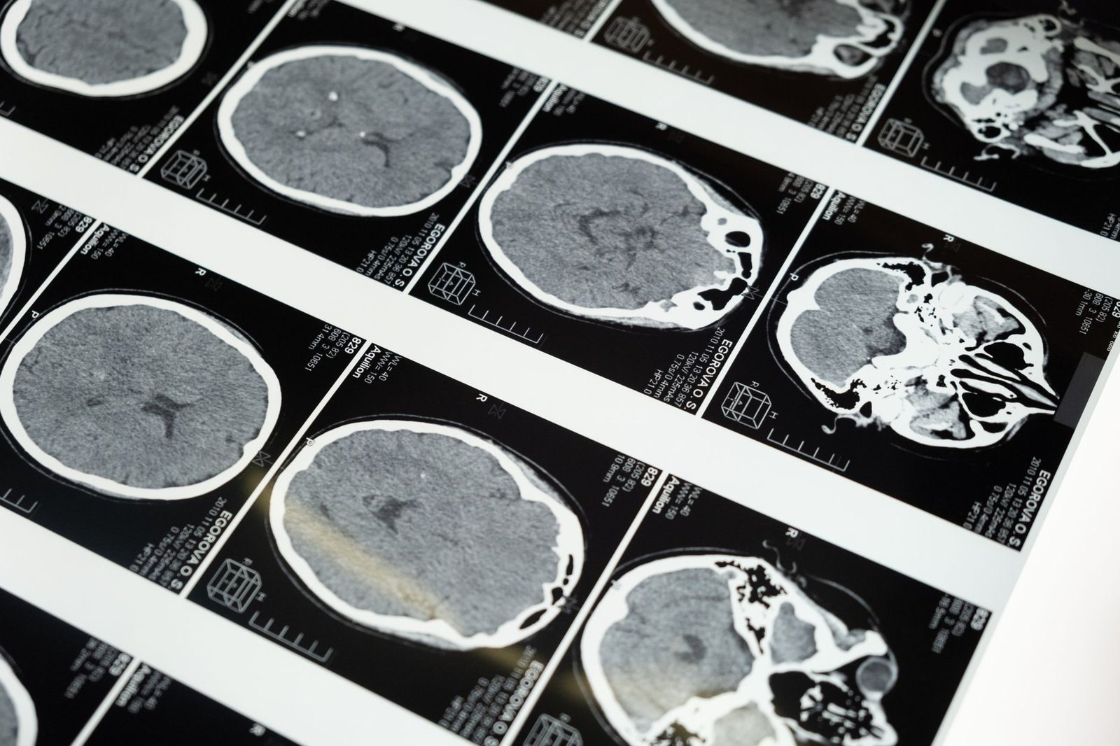
WHY YOUR MRI DOES NOT EXPLAIN IT
Your neck MRI showed degenerative change and a couple of bulging discs. So does the scan of a large share of people your age who have no pain at all.
Is it normal for a neck MRI to show disc bulges with no pain?
Age-related change on cervical imaging is extremely common in people with no neck symptoms whatsoever. That includes disc degeneration, bulges, height loss, facet arthropathy (facet joint disease) and foraminal narrowing (a tighter nerve exit). Its prevalence (how many people have it) climbs steadily with each decade.
That single fact does most of the work on this page. It means a report listing those findings has described a neck that has been used. That is what almost every adult neck is. It has not, by itself, identified your pain source.
Can an MRI miss the cause of neck pain?
It can show a large disc fragment pressing on a specific root in someone whose exam points to exactly that root. It can show cord compression, which is a different order of problem. It can show a tumor, an infection, or a fracture. Those are the findings that truly change management. They are the reason imaging exists.
What it cannot do is tell you whether a facet joint hurts. It cannot tell whether a muscle is referring pain (sending it) into your head. It cannot tell whether a nerve is irritated chemically rather than mechanically. And it cannot tell which of three degenerate levels is the one causing symptoms. Those are not visible. They are answered by examination and, where needed, by a diagnostic block.
The two ways this goes wrong
Over-attribution. A finding gets blamed, treatment follows the finding, and it fails. That is because the finding was an incidental one (there, but not the cause), and the actual pain source was never addressed. In its worst form this ends in an operation on a level that was never symptomatic. That is one recognized route to failed neck surgery.
Dismissal. The scan is clean, so the pain is treated as unexplained or as something the person is doing to themselves. This is common, and it is wrong. Facet-mediated pain, myofascial pain, CRPS and early nerve irritation all cause severe symptoms with entirely normal imaging.
How do you find the pain source when the MRI is normal?
- Map the distribution, because where pain travels narrows the list of structures faster than any image.
- Examine and grade, because objective loss and no objective loss lead to different places.
- Provoke on purpose. If a movement or a palpation (pressing on a spot) brings on your actual complaint, that is evidence the scan cannot supply.
- Test where testing will change something: nerve studies for the nerve question, a diagnostic block for the joint question.
- Then, and only then, read the imaging against the clinical picture, not the other way around.
The order matters more than people expect
Reading the scan first anchors everything after it. Once a report has named a level, the exam tends to be read to fit. Findings that do not fit get quietly discounted. Examining first and reading second is a small discipline in how we work, and it changes conclusions.
It is also why we ask for the images rather than only the report. The report is a reading made without your exam in hand. It was never meant to be the diagnosis.
What are the red flags for neck pain?
Signs that the spinal cord rather than a root is involved — hand clumsiness, dropping things, trouble with buttons and coins, a change in walking, bladder disturbance. Progressive weakness. Fever with neck pain. Significant unexplained weight loss. A history of cancer. Trauma.
Those are on the urgent page, and they do not wait for a sequence of tests. Everything else has time. Using that time on a proper exam is a better investment than using it waiting for a scan slot.
What do the terms on my neck MRI report mean?
Radiology language is descriptive and cautious by design. It reads as alarming to people who are not used to it. Disc desiccation, uncovertebral hypertrophy, endplate change and facet arthropathy all describe ordinary wear. They appear together on most scans of most necks past forty.
If a report has frightened you, that is worth raising directly at the appointment. Going through what each term means against your own exam takes a few minutes. It removes a source of worry that is doing real harm on its own.
What people ask about neck imaging
My report says degenerative disc disease. Is that serious?
It is a description of normal age-related change. It appears on the scans of enormous numbers of people with no symptoms. It is not a disease in the way the phrase suggests. How facet pain is confirmed.
Should I get an MRI before my appointment?
Not necessarily. If there are no red flags, the exam usually decides whether imaging is needed and which imaging. Ordering it first often adds findings that need explaining away. Trigger points and referred muscle pain.
My scan is normal but I am in real pain. What now?
That is a common and entirely explainable situation. Facet joints, muscles and irritated nerves all hurt without showing anything. All three can be tested by other means. The block that settles it.
Why do you want the images and not just the report?
Because the report was written without your exam in front of the radiologist. Reading the two together is a different task from reading either alone. What whiplash actually injures.
Can an MRI miss a pinched nerve in the neck?
Yes, in one important way. An MRI shows a large disc fragment pressing on a root. But it cannot show whether a nerve is irritated chemically rather than squeezed. Early nerve irritation can cause severe symptoms with completely normal imaging. The nerve question is answered by exam and, where testing will change something, by nerve studies.
Can my MRI report tell me what is causing my pain?
Not on its own. A good report describes what is there without claiming what hurts, because the radiologist has not examined you. Say a report names a level as the cause of your symptoms. Then it has gone beyond what the images can support. The diagnosis comes from reading the images against your exam, and it lives where the two overlap.
When does an MRI finding actually explain neck pain?
When the scan and the exam point at the same place. Take a large disc fragment at C6-7 in someone with middle-finger numbness, a weak triceps and a reduced triceps reflex. That is a real diagnosis, and a good one. A C6 finding in someone whose exam shows a C7 pattern is a bystander. The finding earns the blame only when the exam backs it.
Should I have neck surgery because of what my MRI shows?
Not on the scan alone. How sure you need to be should match how permanent the step is. A trial of anti-inflammatory treatment on an uncertain diagnosis costs little. A fusion on an uncertain diagnosis cannot be undone, and the levels above and below it carry the consequence. Operating on a level that was never the pain source is one recognized route to failed neck surgery.
Related reading
- Cervical facet syndrome
- EMG and nerve conduction studies
- Cervical medial branch block
- Trigger points and myofascial pain
- When a neck problem is urgent
- What to expect at your first visit
Have the examination done before the scan decides for you
A scan read against a proper examination is useful. A scan read on its own names findings that most people your age also have.
12174 Natural Bridge Rd, Suite 302
St. Louis, MO 63044
Next to DePaul Hospital, just off the 270 and 70 junction, west of the airport.
Sources
- Kong et al. Prevalence of adjacent segment disease following cervical spine surgery: A PRISMA-compliant systematic review and meta-analysis. Medicine (Baltimore), 2016. PubMed 27399140
- Sheen et al. Cervical medial branch nerve integrity after cervical spine surgery: Considerations for radiofrequency ablation. Interv Pain Med, 2026. PubMed 42180437
- Engle et al. Radiofrequency ablation for the cervical spine. Ann Palliat Med, 2024. PubMed 38902987
- Victory et al. Solitary, asymptomatic, posterior, vertebral, intracanal, cervical spine osteochondroma. Radiol Case Rep, 2011. PubMed 27307940
The two questions worth asking about any scan finding
First: does this finding match my exam? A C6 finding on imaging in somebody with a C7 pattern on exam is a bystander. Second: would treating this finding be reversible? The bar for acting on an unclear finding should rise with how permanent the action is.
A trial of anti-inflammatory treatment on an uncertain diagnosis costs little. A fusion on an uncertain diagnosis cannot be undone. The levels above and below it will carry the consequence for the rest of your life.
What a good imaging report looks like in practice
It describes what is there without asserting what hurts. Say a report names a level as the cause of symptoms. Then it has gone beyond what the images can support, however confidently it is phrased. The radiologist has not examined you and is not claiming to have.
That is not a criticism of radiology. It is a reminder that the report is one input into a diagnosis. The exam is the other one, and the diagnosis lives in the overlap.
When the scan and the examination genuinely agree
Then you have something solid. It is worth saying that clearly, rather than leaving this page sounding like imaging is useless. Take a large lateral disc fragment at C6-7 in somebody with middle-finger numbness, a weak triceps and a reduced triceps reflex. That is a diagnosis, and it is a good one.
The problem is not imaging. The problem is imaging used as a substitute for an exam rather than as a complement to one. That is what happens when the report arrives before anybody has laid hands on the neck.